Comprehensive Retina Care
Advanced diagnosis and treatment for retinal diseases using state-of-the-art technology and expert retinal specialists.
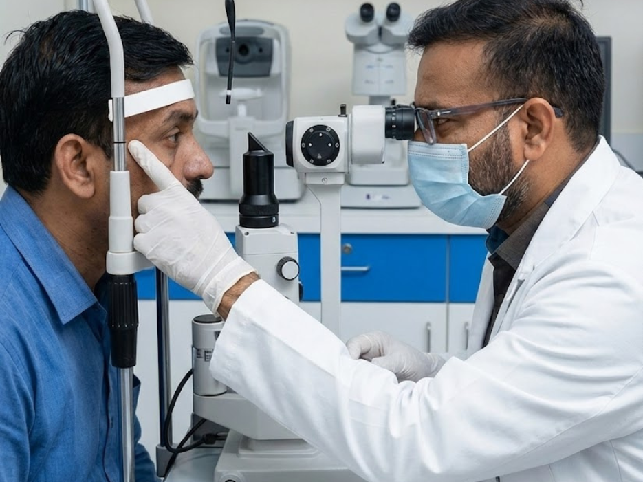
Advanced Retinal Care & Treatment
At Aryavart Eye Hospital, our retina department is equipped with the latest technology for diagnosing and treating complex retinal conditions. Our team of retina specialists provides comprehensive care for macular degeneration, retinal detachments, diabetic retinopathy, and other retinal disorders.
Why Retinal Health is Critical
Vision Processing Center
The retina converts light into neural signals - damage leads to permanent vision loss
Silent Progression
Many retinal diseases progress without symptoms until significant damage occurs
Time-Sensitive Conditions
Conditions like retinal detachment require immediate treatment to save vision
Systemic Health Indicator
Retinal examination can reveal systemic conditions like diabetes and hypertension
Retinal Conditions We Specialize In
Age-Related Macular Degeneration (AMD)
Dry and wet AMD management with anti-VEGF injections and laser therapy
Common in ElderlyDiabetic Retinopathy
Complete management from early screening to advanced laser and surgical treatment
Diabetes RelatedRetinal Detachment
Emergency surgical intervention using vitrectomy and scleral buckling techniques
Surgical EmergencyMacular Hole & Pucker
Microsurgical repair of macular holes and epiretinal membranes
Surgical TreatmentRetinal Vascular Disorders
Treatment of retinal vein occlusions, artery occlusions, and vasculitis
Vascular IssuesUveitis & Inflammatory Diseases
Management of inflammatory conditions affecting retina and uvea
InflammatoryAdvanced Retinal Imaging Technology
OCT (Optical Coherence Tomography)
High-resolution cross-sectional imaging of retina and optic nerve
Fundus Photography
Digital retinal photography for documentation and progression tracking
Fluorescein Angiography
Dye-based imaging to evaluate retinal circulation and vascular abnormalities
Widefield Imaging
Ultra-widefield retinal imaging to visualize peripheral retina pathology
Advanced Retinal Treatments
Vitreo-Retinal Surgical Excellence
Pars Plana Vitrectomy
Micro-incision surgery to remove vitreous gel for various retinal conditions
Scleral Buckling
External support procedure for retinal detachment using silicone bands
Pneumatic Retinopexy
Gas bubble injection for selected retinal detachments
Membrane Peeling
Microsurgical removal of epiretinal membranes and internal limiting membrane
Retina Services FAQs
Common retinal symptoms include:
1. Floaters: Spots, cobwebs, or strings in vision
2. Flashes: Brief sparks or lightning streaks
3. Blurred vision: Central or peripheral blurring
4. Distorted vision: Straight lines appearing wavy
5. Dark areas: Missing parts of visual field
6. Sudden vision loss: Complete or partial loss
7. Color vision changes: Colors appearing washed out
Note: Some retinal conditions like diabetic retinopathy and early AMD may have NO symptoms initially.
Recommended screening frequency:
• Adults 40-54: Every 2-4 years
• Adults 55-64: Every 1-3 years
• Adults 65+: Every 1-2 years
• Diabetics: Annual dilated exam
• High myopia: Annual screening
• Family history of retinal disease: Annual screening
• Previous eye surgery/injury: As recommended
• Existing retinal condition: Every 3-12 months based on severity
More frequent exams may be needed based on individual risk factors.
Pain level: Most patients experience only mild discomfort. We use topical anesthetic drops to numb the eye, and the injection itself takes just seconds. You may feel pressure but typically not pain.
Safety profile: Intravitreal injections are very safe when performed by experienced retina specialists. Serious complications are rare (<1%):
• Endophthalmitis (infection): 0.05%
• Retinal detachment: 0.03%
• Cataract progression: May accelerate
• Temporary increased eye pressure
Benefits vs Risks: For conditions like wet AMD or diabetic macular edema, the vision-preserving benefits far outweigh the minimal risks. Most patients require multiple injections over time.
Vitrectomy recovery timeline:
• First week: Eye patch, limited activity, frequent eye drops
• 2-4 weeks: Gradual return to normal activities, avoid strenuous exercise
• 1-3 months: Vision gradually improves, most activities resumed
• 3-6 months: Final visual outcome apparent
Specific restrictions:
• No heavy lifting (>10kg) for 4-6 weeks
• No swimming for 4-6 weeks
• Avoid eye rubbing
• Positioning requirements if gas bubble used
• Air travel restrictions with gas bubble
Success rates: Modern vitrectomy has >90% success rate for many conditions. Final vision depends on preoperative condition and duration.
It depends on the condition and timing:
Reversible conditions:
• Macular edema (swelling)
• Some retinal detachments (if treated early)
• Vitreous hemorrhage (blood clears)
• Central serous retinopathy (often resolves)
Partially reversible:
• Wet AMD (stabilize, some improvement)
• Diabetic retinopathy (prevent progression)
• Retinal vein occlusion (limited improvement)
Irreversible damage:
• Photoreceptor cell death
• Long-standing retinal detachment
• Advanced geographic atrophy (dry AMD)
• Optic nerve damage from glaucoma
Key point: While we cannot regenerate dead retinal cells, we can often prevent further damage and preserve remaining vision. Early intervention is crucial for best outcomes.